
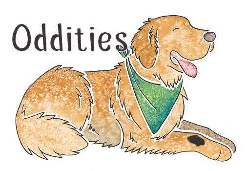
Oddities
Already know what you're looking for? Quick Links | ||
Somatic Mutations and Chimera
A somatic mutation is a mutation that occurs in the body cells after the embryo has begun to form. Cells are divided into two categories - germline cells (i.e. gametes - sperm and eggs) and somatic cells (all other body cells). When a mutation occurs at the point of conception (when the first cell begins to replicate) then this mutation may enter the germline and so be passed on to future generations, but after this point the two types of cell replicate separately and any mutations occuring in the somatic cells will only be passed on to the descendents of those cells and will not be passed to the next generation. This is because only the information carried in the gametes will go on to form a new individual (of course, mutations may well happen in the gametes too during the individual's lifetime, and these mutations will be passed on).
This may sound complicated, so perhaps an example will make it clearer. Cancer is a somatic mutation. Sometimes a mutation occurs in a cell that makes the cell "immortal". Most cells can only replicate a certain amount of times, but when this particular mutation happens the cell is able to replicate itself indefinitely (this is the biological definition of immortal), and this is how tumours form and also why they can be very difficult to get rid of. However, although the predisposition to cancer may be genetic, cancer itself is not as it occurs as a mutation in cells that will not go on to form new individuals.
If a pigment somatic mutation takes place during the development of the embryo, patches of another colour may appear on the animal. This is often seen as black patches on recessive red Labradors and Golden Retrievers. The painting at the top of the page shows what this might look like. It is not certain why pigment mutations seem to appear in these breeds more than others, however it's possible that the recessive red gene is unstable and particularly prone to mutation. All it takes is for one cell to mutate early on in development - then all cells descended from that one as the embryo grows will be the different colour. Due to the mutation these patches effectively have a different genotype to the rest of the dog, and this is sometimes known as mosaicism.
Sometimes you will see a colour test result on a dog that appears to show three or more alleles on a locus. This is particularly common on merles, and also sometimes seen on the B locus. This is due to mosaicism. It is generally visible on merles (they will often have large areas of solid colour), but on colours such as liver/brown, the different b alleles express the same and you won't be able to tell from looking at the dog that they are a mosaic.
A very rare form of mosaicism is the chimera. A chimera is a single animal that is the combination of two separate embryos. You may have heard this term used to describe creatures with the DNA of two different species spliced together, such as human/mouse embryos created in labs, but it also occurs naturally when two fertilised eggs combine very early in development. Effectively, the resulting animal is two individuals in one. It has patches of one set of DNA and patches of the other as well. Sometimes it can be impossible to tell a chimera without genetic testing (particularly in humans), but in some cases the resulting animal may be a combination of two different colours. I do not know of any proven dog chimeras, but the effect may be similar to the somatic mutations already discussed, but more extensive (roughly 50/50 of each colour). Chimaeras in some species can produce a brindle-like pattern.
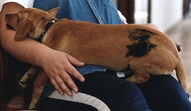
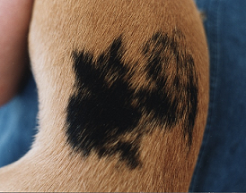
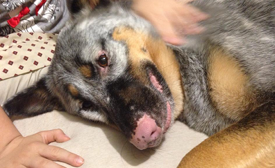
These pictures were submitted by Dr Anna Laukner and show a small dog from a village in Ibiza with a somatic mutation. Black patches on a red base is the most commonly seen somatic mutation. If the black on this dog was due to the sable gene, it would be much more evenly distributed through the coat.
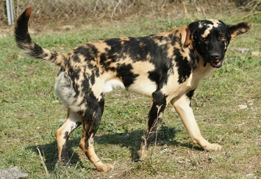
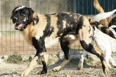
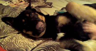
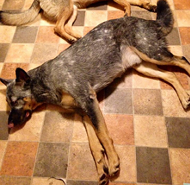
This incredible Labrador appears to show somatic mutations, but is a possible chimera due to the extent of his patching. These photos were taken at a rescue centre in France and sent to me by Damien Ricco but
we have been unable to locate the original photographer. If you took these photos then please contact me and I will be happy to add credit (or remove them if you wish).

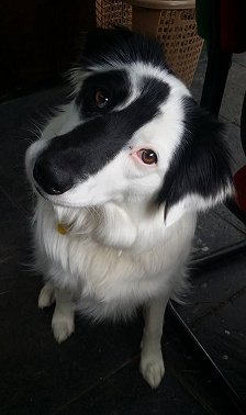
Maza's striking face markings could be a somatic mutation or may just be unusual piebald markings. They appear to follow the opposite pattern to the usual face markings seen on dogs with white spotting. The beautiful Maza is owned by Jenny Ban.
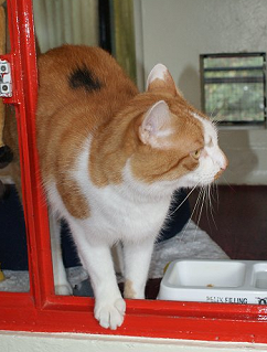
A somatic mutation on a ginger cat.
Vitiligo
Vitiligo is a condition where the skin/coat cells stop being able to produce pigment over the course of months or years, causing areas of white. It is a catch-all term for a variety of conditions with lots of different causes, both genetic and non-genetic (including an auto immune disease that attacks pigment cells), but in dogs does not usually have any adverse effects other than coat colour. The pigment loss is generally concentrated around the head/face, but often spreads to the rest of the body.
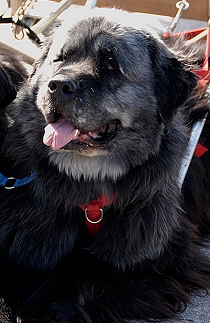
Phaergus, the Newfoundland above, has developed what appears to be vitiligo on his head, chest, back and tail. The first picture shows Phaergus in May 2010 (at 3 years old), and the second shows him in March 2011. Phaergus and his photos belong to Bobbi Walker.

Sarge the German Shepherd Dog, see: Sarge Flufferson on Facebook
Sarge here shows the progression of extensive vitiligo. As you can see in the first and second pictures, he started as a black-and-tan and his nose pigment seems to have been the first thing to go. He has then developed almost a roaning pattern of white hairs on his black areas, becoming more and more white over time. Unlike Phaergus above, Sarge's vitiligo is all over his body and has affected his nose, lip and eye rim pigment, suggesting it has a different cause to Phaergus's vitiligo. Interestingly, Sarge's condition only affects his black areas (the tan areas have no white hairs at all), and the black mask on his muzzle has not been affected to the same extent as the rest of his black. This could provide a clue as to the cause of Sarge's vitiligo.
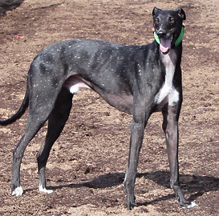
"Snowflake" Greyhound Spots
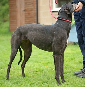
This phenomenon appears to occur fairly frequently in black Greyhounds from certain lines. There are small white spots on the coat, almost like ticking in reverse. These dogs look like they have been sprinkled with snow or white paint. It is not clear whether this is caused by some form of greying, vitiligo, or whether it is actually a white patterning gene. Vitiligo is unlikely as this usually becomes more extensive over time, as with Sarge the German Shepherd Dog in the section above.
Similar markings sometimes occur in horses and are known as "birdcatcher" spots.
Nick Irish, owned by Dr Tess Tsindos.
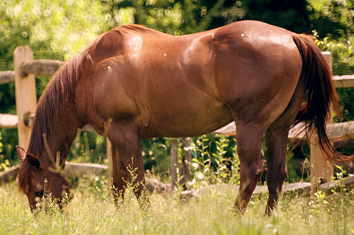
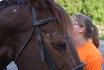
Rambo the Quarter horse, submitted by Andrea de Carlo, shows birdcatcher spots on both his head and body.
Fever Coats
Sometimes litters are reported where the puppies are a light grey colour despite not being dilutes. These puppies usually turn black within a few weeks or months as the initial coat moults out. This phenomenon is known as "fever coat", and is more commonly reported in kittens. Fever coats are generally caused by a minor illness or stress in the mother very early in development, which affects the production of pigment in the foetuses. Most cases of fever coat in dogs have been reported in Border Collies and Bearded Collies.

** Please note that I am not a research scientist, and the information on this page comes from my own knowledge and observation of dogs, observational and testing data provided via e-mail by site visitors, any research papers linked on the page, and the information provided by Dr Sheila M. Schmutz on her excellent website http://homepage.usask.ca/~schmutz/dogcolors.html
For further genetics resources, see the Links page