Already know what you're looking for? Quick Links | |
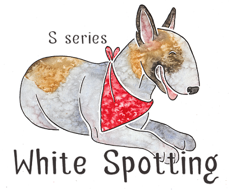
The White Spotting Series
Most white spotting on dogs is determined by the genes on the S locus. When we use the term "white spotting" we simply mean
white areas on the dog, not actually white spots. White spotting can occur on any colour, and will cover up both eumelanin and phaeomelanin. In technical terms this is
known as epistasis. So any dog
can have white markings, whether they're black, blue, liver, isabella, brindle, sable, tan-pointed, merle or whatever.
White hair occurs when the skin cells are unable to produce any pigment. The white spotting gene impairs the ability
of cells on particular parts of the skin to make pigment, so the skin becomes pink and the fur white. Nails and paw pads will also become pink in areas
where pigment is not produced.
So far, only two white alleles have been proven to exist on the S locus:
A third allele may exist for "extreme white" (sw), however this has not been proven and so far all dogs with high white have been
shown to be homozygous for sp instead.
The white spotting alleles are thought to be examples of incomplete dominance. This means that a heterozygous dog will express
its most dominant gene, but may also be affected by the more recessive one to a lesser extent. For example an Ssp dog may have some white spotting (see below). However,
the relationship between the alleles is complicated and can vary between breeds.
It has been shown that some dogs with white spotting do not have an sp allele at all. These are mostly dogs with "true" irish spotting (in other words, irish spotting that breeds true - this should be made clear further down the page). The allele
that causes this pattern has not yet been identified and it is not known if it is also located on the S locus. For the purposes of this site we will refer to this gene as
si, but remember that it is most likely located on a different locus to sp. si is much less common than sp and only occurs in certain breeds.
Spread of White
Whichever white pattern a dog has, its white will always follow the same rules of spread. White starts on the farthest "edges" of the dog - the tail tip,
the tip of the muzzle, the paws and the tip of the breastbone. This is known as the "trim" pattern. From there it spreads to cover the muzzle and forehead, the front of the chest, the lower legs and more of the tailtip, creating
irish spotting.
Next it spreads round from the front to the back of the neck, and creeps up the legs and tail. On a piebald dog, only the head, back and tail base may still
be coloured. The back colouring is the next to go, followed by the tail base, then the face markings. The ears will always remain coloured unless the dog has a very
high amount of white. The ears are generally the last part of the dog to turn white.
Of course the idea of white "spreading" is metaphorical, to give you a picture of how white patterning works. White
doesn't spread like this on one particular dog (i.e. you won't get a solid coloured puppy that gradually loses colour as it grows, until it's almost white! Although puppies do often lose or gain a little
colour as they grow), it's just to show which areas remain coloured on dogs with more and more white. One way to
think of it is that the dog retains colour best in the most important areas of its body - around its internal organs (body and tail base patches) and its brain (ears and face patches) - and
loses colour easiest from the parts farthest from these areas. In technical terms, pigment "migrates" to different parts of the body during the development of the embryo, and the S gene determines how far
the pigment migrates. Sometimes it simply doesn't reach the furthest extremities (this can be caused by a minor problem or illness during development), and this can result in a small amount of white trim
on a dog without sp, for example a small chest patch on an otherwise solid-coloured dog.
The white rules aren't set in stone - sometimes individual dogs can have unusual white patterns, where, for example, the white on the
legs is very uneven, or they have piebald patches in unexpected places, like on the neck or chest. However, in general, they do hold relatively true.
Residual White and White Trim
A very small amount of white on the chest, toes or tail may occur when the pigment doesn't migrate fully as the embryo develops. This is known
as residual white and can sometimes be caused by minor illness in the mother or in the embryo, or may have no obvious cause at all. There is no particular genetic basis for residual white, however it's clear that some dogs have a predisposition to it which can be passed on to their offspring.
If a slightly larger amount of white is present then the dog may be heterozygous for sp, in other words Ssp. In a breed
such as the Newfoundland you would get such a dog from crossing a "Landseer" (piebald, spsp) with a solid (SS). However, in breeds carrying piebald there is no real way to know whether minimal white markings are just residual white
or indicate the presence of the piebald gene without genetic testing or test breeding, as piebald heterozygotes may have anything from a tiny chest spot to pseudo-irish markings (see below).
Irish Spotting Pattern
Irish spotting (si) is the pattern sometimes known as "boston" or "mantle", although these terms do not always refer to "true" irish spotting. On a dog with irish spotting, white is found on the legs, the
tip of the tail, the chest, neck and muzzle. Many dogs with this pattern have a full white neck ring and a blaze.
True irish spotting is caused by an as yet unidentified gene, but we can assume irish spotted dogs to be homozygous for it (sisi) as it breeds true. This means that two irish spotted dogs bred together will produce puppies with irish spotting, and the white will not increase. We can assume that a solid dog bred to
an irish spotted dog will produce a heterozygous dog with less white (a white trim, as shown in the section above).
The term "irish spotting" actually comes from a term used in the early 20th century to describe a white pattern found in rats in Ireland.
Pseudo-Irish
"Pseudo" irish spotting may look the same or very similar to true irish spotting, but is in fact not caused by sisi but by Ssp, i.e. these dogs are really heterozyous or potentially even homozygous piebalds. The incomplete dominance of S means that an Ssp dog may show up to
roughly half the amount of white as an spsp dog. These dogs do not breed true and when two are crossed
the puppies may be solid, piebald or inbetween. See below for an example of this in Boxers.
Note that not all Ssp dogs show much white, or in some cases any white at all. The amount of white on a piebald heterozygote appears to vary drastically and some may look exactly like homozygous solids.
Finally, a "flashy" irish spotted dog (one with more white than usual) may be caused by a combination of si and sp. If a true irish spotted dog
also carries an sp allele then the normal white pattern may be extended. This supports the theory that si is actually on a different locus, as the two alleles appear to
be inherited completely separately. This has been shown to occur in Shelties, where dogs carrying the sp allele as well as irish spotting can usually be identified by having more
white around the neck and underside of the body. An spsp Sheltie has a high amount of white and is known as a "colour-headed white". Shelties are one breed known to carry both true irish spotting and the sp allele, but many breeds only have one or the other.
Piebald Pattern
Piebald (spsp) usually produces a coloured head (with or without white on the muzzle and as a blaze), and patches
on the body. Generally the base of the tail is coloured, but other than that the patches may be located anywhere on the body (but rarely on the legs).
Extreme White Pattern
The extreme white pattern consists of a completely or predominantly white dog with just small amounts of colour on its head and sometimes base of tail. Small body
patches may be present too. Sometimes the nose is pink or partly pink, and the eyes may be blue in some breeds due to lack of pigment.
So far all extreme white dogs that have undergone genetic testing have been shown to be homozygous for the piebald gene (spsp), just like the piebalds
in the section above. However, as there is a fairly large difference between those dogs and the ones shown below, it is possible there is something else going on to cause the high white. In breeds with both true irish spotting and piebald the high white may simply be caused
by the interaction between homozygous irish spotting and homozygous piebald (e.g. the Sheltie). In other breeds the cause is less obvious and has led some people to postulate a further S allele - sw. However, no evidence has yet been found for the existence of sw, on the S locus at least.
Extreme white can occasionally cause problems when it removes large amounts of pigment from the face and ears. The most common
problem is deafness (due to lack of pigment in certain parts of the inner ear, which prevents it from functioning properly), but dogs with exposed unpigmented (pink) skin are also more prone to skin cancer than those with more pigment.
Split Faces and White Heads
There is thought to be a separate gene or modifier that causes some dogs with the irish spotting, piebald or trim pattern to have a split or completely white face, even if there's very little other white on the dog.
A split face is when half of the face is white and the other half is coloured.
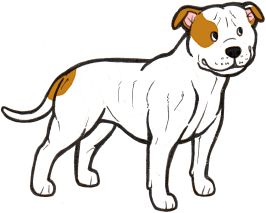
Split and completely white faces are particularly common in the bull-type breeds, e.g. English Bulldog and Staffordshire Bull Terrier. The cause is unknown, however it's interesting to note that many "panda" German Shepherds (see below), which are known to have a different white mutation to most other breeds, have split faces in a similar pattern, so it's possible that split/white faces could be caused by the same or a similar gene.
Ticking and Roan
Any white areas on a dog, no matter how big or small, may be ticked or roaned due to the T gene. The ticking corresponds to the colour the area of the coat would have been if it wasn't white, which means that it can vary across the body. For example, a black dog with tan points will have black/grey ticking where it would have been black, and red/tan ticking where the points would have been. See the Ticking page for more information.
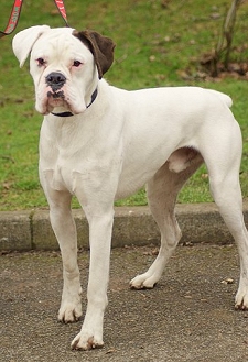
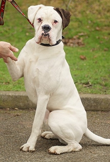
White Boxers
Boxers generally come in what appears to be the irish spotting pattern, so
we would expect most examples of the breed to have sisi on the S locus. However, sometimes Boxer puppies are born which
are completely or almost completely white. How these puppies could be regularly born to parents with much more colour perplexed Boxer breeders for
a long time.
However, we can now provide an answer to this. Boxers do not have the si allele, and supposedly irish spotted Boxers are actually pseudo-irish - i.e. Ssp. When two pseudo-irish dogs are bred together
some of the puppies will be homozygous piebalds (spsp). Boxers with no piebald allele can still have residual white on the extremities, and pseudo-irish dogs can have anything from a very small amount of white to full irish markings, which can sometimes make it difficult to distinguish a piebald carrier from a non-carrier.
White puppies can therefore be mostly avoided by always breeding irish-marked dogs to solid dogs, although care must be taken when it is unclear whether a dog is genetically solid or pseudo-irish, as it is possible for solid dogs to carry a piebald allele and not express it at all or only partly.
White Boxers come with many of the problems associated with high-white dogs, including a high rate of deafness.
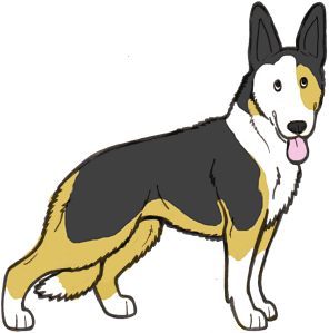
"Panda" Shepherds
So called "panda" German Shepherd Dogs, like the one pictured, have been found to have a completely different white mutation to normal dogs with this pattern. Although these dogs look like they have irish spotting, the pattern is actually caused by a different gene entirely, known as "KIT". "Panda" is a dominant mutation, and like many white patterns caused by the KIT gene in other species, it is an embryonic lethal, which means that when an embryo has two copies of the gene it will be reabsorbed into the womb. Heterozygous dogs have no known health problems linked to the gene, however. Harlequin in Great Danes is another example of an embryonic lethal in dogs, although caused by a different gene to panda.
Some panda Shepherds have blue eyes, however this is not linked to the KIT gene. Another observation is that many panda Shepherds have split faces and very few seem to have the neat blazes often associated with true irish spotting or the piebald gene.
A few similar spontaneous white mutations in other breeds have been shown to have been caused by the same gene, including a mutation in Weimaraners. However, these mutations are very rare and the German Shepherd seems to be the only breed where a KIT mutation has become established.
"False" Whites
Sometimes white can occur on dogs separately to the S locus white spotting. One example is as part of the double merle pattern. A double
merle will almost always have more white than its parents, and will often appear to have the piebald or extreme white pattern when in fact it does not
carry those alleles. The harlequin gene also causes a similar effect. See the Merle page for more information.
White can also occur due to dilution of phaeomelanin by the I locus. Phaeomelanin is red pigment, and the I locus
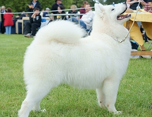
can dilute it to cream, ivory or sometimes even white. Breeds such as the Samoyed have this second
type of dilution, so they appear completely white but in fact it's not due to white spotting. They are recessive red (so they cannot
produce any black pigment) with dilution of their red pigment to white, resulting in a solid white dog with black nose pigment. They also test as homozygous for recessive black, but it seems the recessive red overrides this.
The main way to tell a dog with extreme white spotting apart from a dog with phaeomelanin dilution is
to look at the pigment on the nose, lips and eyerims. A dog with extreme white spotting is likely to be missing some pigment in these areas, so
they will be partly or completely pink. A dog with phaeomelanin dilution will have solid black in all these areas (possibly with a dudley nose, which
are common on dogs with dilution -
see the Nose Colours page).
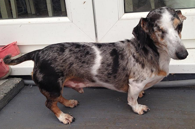
These two Dachshunds look just like piebalds, but they're actually double merles and most likely don't have any S-locus white markings at all. The pattern produced by double merle is strikingly similar to a homozygous piebald, although it can sometimes be less regular and extreme split faces going all the way down the muzzle are very common (as on the second dog).
Quick Summary!
The S locus in dogs has two known alleles: S (no white markings), and sp (piebald). Due to incomplete dominance, one copy of the piebald allele results in a dog with minor white markings (often called the "trim" pattern), and two copies causes piebald or extreme white. A very wide range of patterns is caused by sp.
In addition to sp, there is another allele known as si (irish spotting), which is most likely located on a different locus. A dog with one copy will have a white trim and a dog with two copies will have full irish spotting (white neck/collar, face, chest, legs and tail tip). The combination of si alleles and sp alleles is thought to cause most of the range of white markings found in dogs.
Recently a rare mutation has been found in another gene known as KIT, and this causes the white markings on "panda" German Shepherd Dogs.
Further Info and Links
The gene causing the majority of white markings in dogs is known as MITF (Microphtalmia-Associated Transcription Factor). This gene causes white markings in a number of mammals and is often associated with blue eyes and deafness. The link between MITF and eye colour and hearing in dogs seems to be weaker than in some other species, although many high-white breeds such as Bull Terriers and Dalmatians do suffer from high rates of deafness. Interestingly, mutations in MITF have no link to skin colour in humans but do cause eye and sight issues.
In other species, some white markings are also caused by the KIT gene. KIT is an extremely important gene and plays a role in stem cells and in the digestive tract, and some mutations in this gene (not those associated with white markings!) can also be associated with a number of different cancers. Most mutations in KIT causing white markings have no associated health problems when heterozygous, but are embryonic lethals when homozygous. KIT is responsible for "black-eyed white" phenotypes in many species, and has no association with either blue eyes or deafness. In dogs, KIT mutations have so far been confirmed in "panda" German Shepherds and in Weimaraners, but are not thought to be widespread.
The other main gene causing white spotting in other species is EDNRB (Endothelin Receptor Type B). No mutations in this gene have been found in dogs, however in horses EDNRB causes "lethal whites" (overo lethal white syndrome), where homozygous foals do not have a fully functioning digestive system and die soon after birth. A number of human diseases are also associated with EDNRB.
Links to studies:
** Please note that I am not a research scientist, and the information on this page comes from my own knowledge and observation of dogs, observational and testing data provided via e-mail by site visitors, any research papers linked on the page, and the information provided by Dr Sheila M. Schmutz on her excellent website http://homepage.usask.ca/~schmutz/dogcolors.html
For further genetics resources, see the Links page
S - no or very minor white
sp - piebald
Of course this means that technically, white doesn't spread at all - it's actually the colour that spreads.
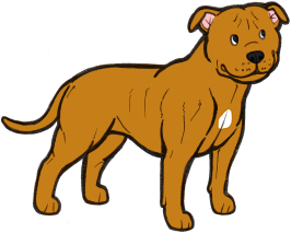

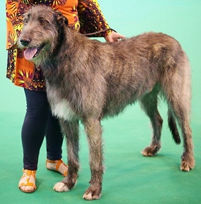
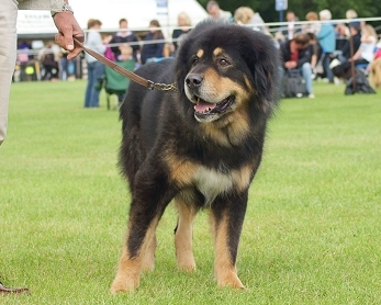
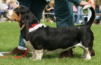
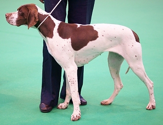
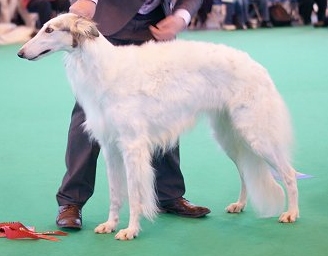
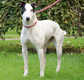
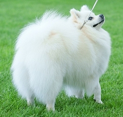
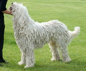
We can assume that the two dogs above are SS and that their markings are just residual white. This is because neither breed comes in piebald or irish spotting. If either of these dogs did have an sp or si gene
then we would expect to see dogs with much more white being produced in these breeds, but this doesn't happen. As it is, their white is non-genetic and breeding two dogs with white markings in these breeds will not necessarily produce puppies with any white at all.
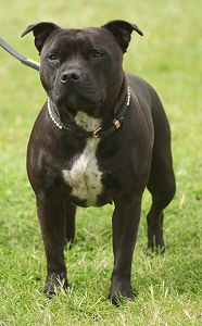
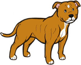
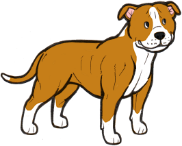
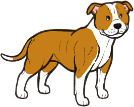
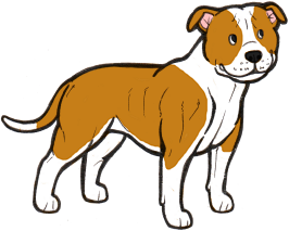
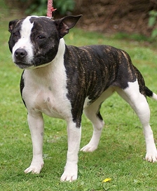
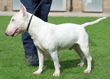
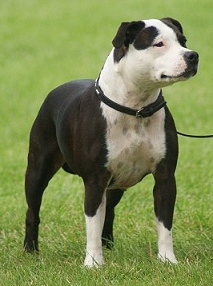
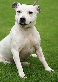
This Staffordshire Bull Terrier is a possible piebald heterozygote (i.e. carrier of the piebald allele). We can't know for sure, but this is the most likely explanation for its white chest patch as the Staffie
breed is known to commonly have the piebald gene. If this dog were bred to another sp carrier then some of the puppies may be piebalds and have much more white than either of their parents.
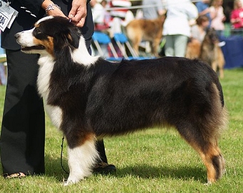
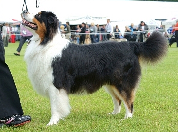
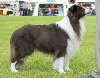
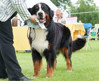
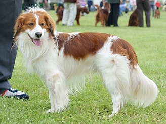
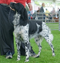
The Aussies, Border Collie and Bernese Mountain Dog shown here are all true irish spotted. None of these breeds regularly come in piebald or extreme white and their white markings breed true (implying they are homozygotes).
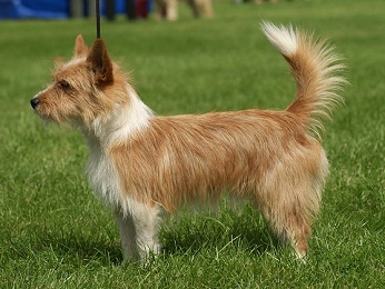
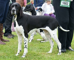
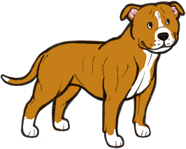
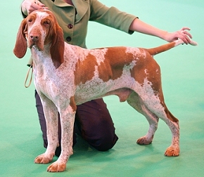
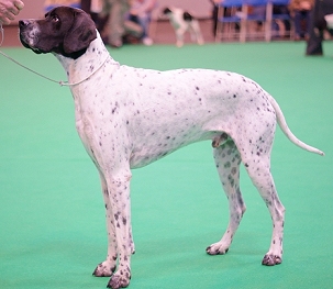
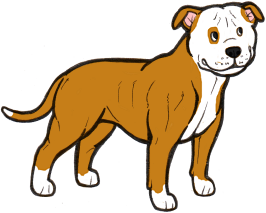
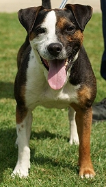
The three breeds above (Staffie, Podengo Portugueso and English Pointer) all carry piebald but are not known to carry irish spotting, so these dogs are most likely pseudo-irish. A true irish spotted dog will not
usually have white on the hips/knees or underside of the body, so this is another clue that sp is present.
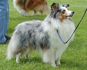
This irish spotted merle Sheltie has a large amount of white and may be a piebald carrier. Piebald carriers are often referred to as "white-factored" and are generally identified by having white extending further up the hind legs (onto the knees).

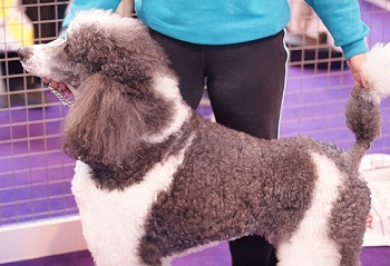
Because piebald is a recessive gene and heterozygotes (piebald carriers) don't always have any white markings, it can remain hidden and
pop up unexpectedly. Both the Poodle (as shown here) and the Shar Pei, traditionally solid-coloured breeds, occasionally produce piebald (known as "flowered" in Shar Pei).

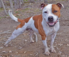
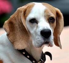
Saxon the Staffie submitted by Christina Jacobs (see Saxon's Facebook page here), and Beagle submitted by Viktoria Kastner
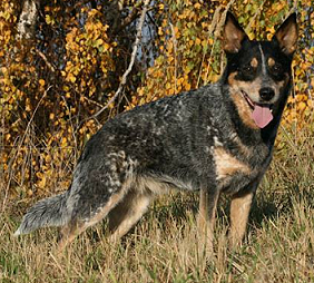
Australian Cattle Dog photo submitted by Viktoria Kastner
Believe it or not, the Australian Cattle Dog above is an extreme white piebald. The solid black on the head is the actual markings, and the solid appearance of the rest of the
coat is created by very heavy roaning, including the tan points. This dog will have been born white with colour on the head only, as roaning develops later. The Large Munsterlander to the right shows heavy ticking on a piebald dog. Ticking is generally lighter than roaning, and the individual spots may be larger.
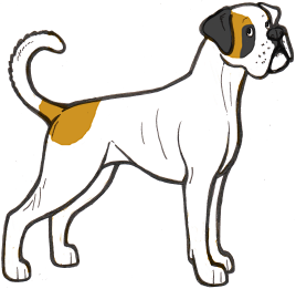
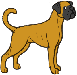
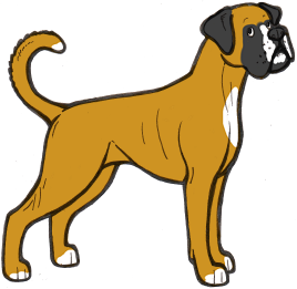
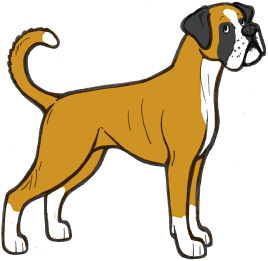
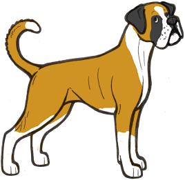
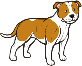
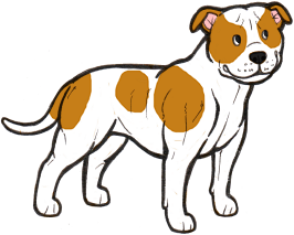
The illustrations above show the range of white markings in Boxers. It is likely that the sp allele here is affected by a modifier that extends the white markings, as white boxers are always extreme white instead of normal piebald pattern.
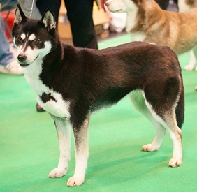

These two dogs (a Siberian Husky and a Finnish Lapphund) are genetically black and tan (atat), but with dilution
of their tan points to white, most likely to due the Northern domino (Ed) gene. It can be easy to mistake diluted points on a domino or black-and-tan dog for white markings, but points will generally be in a very regular and symmetrical pattern. The Husky has
actual white spotting as well - note the irregular pattern on the chest and the thin blaze on the muzzle.
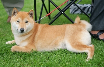
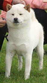
Urajiro is another phaeomelanin dilution pattern that can look like white markings. Urajiro is when the dilution is confined to the "points" of the dog, like on the Shiba Inu here. See the Phaeomelanin Dilution page for more information.
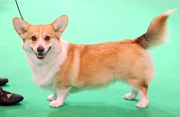
Some dogs can have both phaeomelanin dilution and white spotting, like the Pembroke Welsh Corgi pictured above. This dog has the white collar associated with irish spotting, but also the symmetrical cheeks associated with urajiro. If you look closely, you can see that the cheeks are an "off-white" colour, not bright white like the collar.

First photo provided by Tina West, second by Dee Allison
One of these dogs is not like the others . . . but which is it? All are "false" whites except for one, which is an extreme white piebald. If you guessed
the Staffie, you'd be right. She has pink around her eyes, ears, muzzle and underside (a sign of lack of pigment, associated with extreme whites) and a few dark spots on her ears.
All the other dogs are recessive reds (ee) or clear sables with phaeomelanin dilution. Note the slight cream sheen on the coat
of the German Spitz, Samoyed and Shiba, and the jet black lip and eye rim pigment on all of them. The Shiba has a dudley nose, often associated with recessive red.
No time to read the whole thing? Here's the quick version!
MITF and White Spotting in Dogs: A Population Study: http://jhered.oxfordjournals.org/content/100/suppl_1/S66.full
A Simple Repeat Polymorphism in the MITF-M Promoter Is a Key Regulator of White Spotting in Dogs: http://journals.plos.org/plosone/article?id=10.1371/journal.pone.0104363
A de novo mutation in KIT causes white spotting in a subpopulation of German Shepherd dogs: http://www.ncbi.nlm.nih.gov/pubmed/23134432
Exclusion of EDNRB and KIT as the basis for white spotting in Border Collies: http://genomebiology.com/content/1/2/RESEARCH0004
Spotted Weimaraner: KIT Gene Strikes Again: http://colorgenetics.info/canine/spotted-weimaraner-kit-gene-strikes-again#sthash.XjGhqSlH.dpbs